SEM in the Spotlight:
Biology of Lichens comes to the Scanning Electron Microscope (SEM) Lab
Ben Zink, Graduate Student, EFB
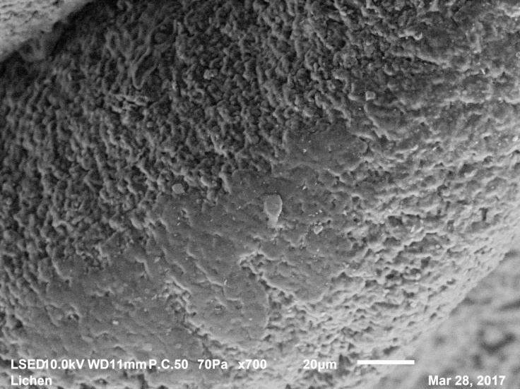
The new JEOL JSM-IT100 SEM at ESF offers many advantages over the previous, outdated model. With ESF being a university with the majority of its research environmentally and biologically related, a main advantage of the new microscope comes with its low-vacuum mode. This method of imaging allows the user to capture images of unfixed, uncoated specimens and is a major advantage when trying to image delicate biological specimens. The low-vacuum mode was demonstrated this semester to the EFB 496 class Biology of Lichens, taught by Dr. Alex Weir. Dr. Weir traditionally brings mycology related courses to the SEM lab as an introduction to electron microscopy and is an advantageous way to bring recognition to the NC-Brown Center. Lichens, being a fragile combination of algae and fungi, were a perfect candidate for a demonstration utilizing the low-vacuum mode. The following micrographs were captured from the lichen samples observed by the students during the demo.


Leave a comment